


Useful
Computer tomography at DENTA VITA clinic

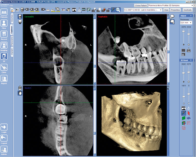
Cone-Ray Computer Tomography (CRCT) is one of the methods of studying bone tissue condition, which is based on radiation diagnostics. Yet, in comparison with traditional medical tomography, the radiation load is significantly smaller, thanks to the cone-ray-intermittent scanning method (the total scan time takes about 6 seconds). Along with orthopantomogram as well as precise X-ray, it is actively used in dentistry, implantology as well as otorhinolaryngology. It is quite different from traditional method of two-dimensional X-Rays in that it allows us to get not merely a flat 2D, but a three-dimensional 3D image of the entire jaw along with the maxillary sinuses.
Computed tomography: indications for usage
- bone volume evaluation
- bone tissues condition and quality assessment
- three-dimensional study of tissues diseases or neoplasms (cysts, granulomas, etc.)
- volumetric study of maxillary and frontal sinuses
- examination of retina (uncut) teeth position
- detection of pathological gingival pockets along with their volume
- identification of additional root canal channels, root form, etc.
Computer tomography: advantages
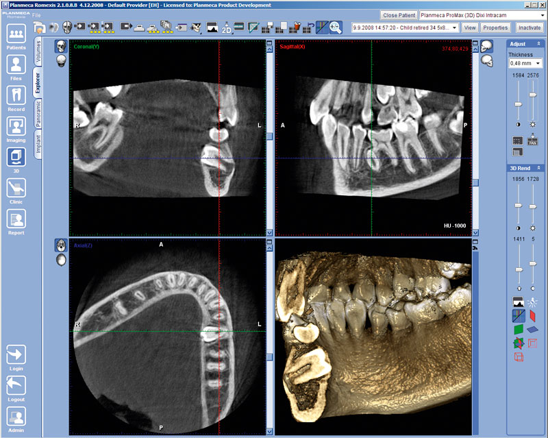
- three-dimensional image: tomography scans the object from several sides, which is why the bone tissue can be viewed in a three-dimensional image (height, width, thickness)
- the thickness of the tissue layer that we see on the screen is only 0.2 mm, and for any kind of radiography, this is the sum of all the shadows of the tissues obtained when the X-ray passes to the sensor or film, so in case of computer tomography the image is clear and does not contain any unwanted shadows
- keeping the result: you can regain the results if necessary, since they are all stored in the clinic's database
- no distortion: the image is accurate - 1:1, not smeared and not stretched as it could be in case of using an ordinary radiograph
- rapid screening and quick appearance on computer display
- safety: the radiation load during the tomography (CRCT) is minimal (just as it in X-ray examination) and the amount of information received is maximal!!!
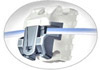
CRCT at DENTA VITA clinic
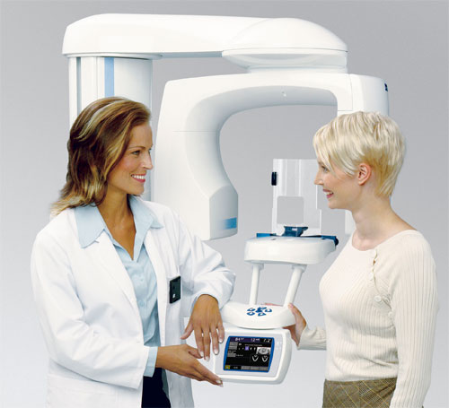
Our center has installed the most innovative equipment, which meets the highest world standards, significantly reducing the radiation dose received by the patient during the research, since it has digital sensors that are very sensitive to X-rays and therefore do not need a high level of radiation!!!
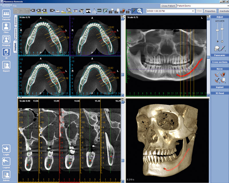
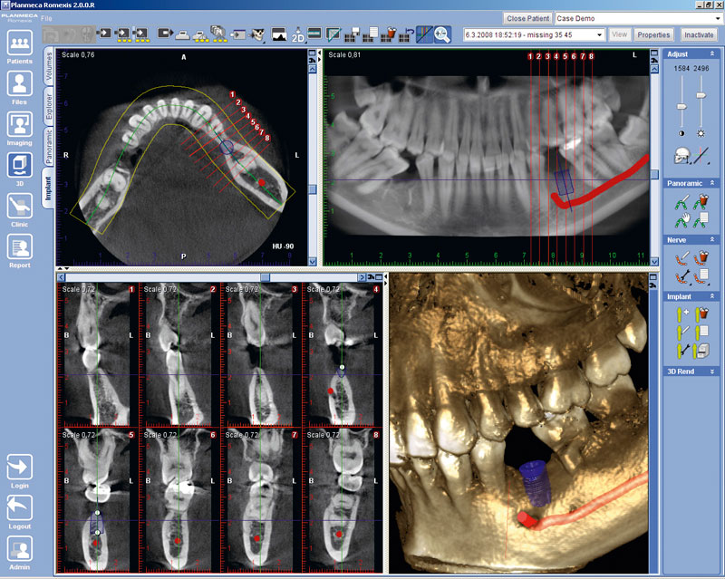
ProMax 3D (Finland) dental tomography allows conducting detailed studies of bone tissue, root of the teeth, temporomandibular joint, nasopharynx, maxillary sinuses, as well as the upper spine. The equipment distinguishes a wide range of areas for scanning - from minimal to study any kind of local problems, to maximal - for diagnosing the condition of large tissue volumes, for instance, the condition of both jaws and joints of the patient.
Close cooperation with 3D-DIAGNOSTIC - the best diagnostic center (www.3d-diagnostic.md) allowed us to experience diagnostics in a new way.