


- Anesthesia
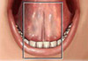
- Kofferdam
- Prophylaxis
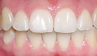
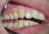
- We treat Teeth
- We treat gums
- We make dentures
-
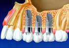
We implant
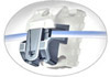
- The laser in implantation
- Kinds of tooth implants
- Implantation stages
- Advantages of implants
- Care of implants
- Service life of tooth implants
- We create conditions for implantation
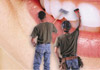
- We operate (surgery)
- We align teeth (orthodontics)
- We treat teeth with children
- We treat with LASER


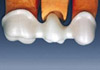
We implant
We create conditions for implantation
Sinus-lifting
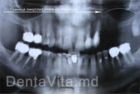 Border of sinus-lifting before operation
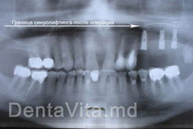 Border of sinus-lifting after operation
Sinus-lifting represents a surgery meant to improve the conditions for placing dental implants in cases of atrophy of upper jawbone.
There are 2 types of surgeries sinus-lifting: open and closed). Closed "sinus-lifting" is performed when bony tissue of upper maxillary sinuses are not less than 10 mm. But if this distance is less than 10 mm, then the "sinus-lifting" surgery will be open.
When is the "sinus-lifting" surgery performed?
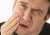
"Sinus-lifting" surgery is recommended in cases when lateral parts of upper jawbone are atrophied. Porous bone of upper jawbone strongly differentiates from the bone in other parts of the body - it has large spaces and initially a little volume of bony tissue. When teeth are lost, pneumatization of maxillary sinus takes place and the quality of bony tissue is getting worse.
In case if bony massif increases in sinus region then "sinus-lifting" allows the surgeon-dentist using longer implants, which is preferable in lateral sides of jawbone in order to create conditions for an adequate resistance occlusion pressure. According to the thickness of alveolar apophysis, dental implants can be placed either simultaneously while performing "sinus-lifting", or in a while after wound healing.
A professional execution of "sinus-lifting" reduces to minimum the complications rate among patients.
Creating conditions for implantation
When a bone is not enough big for implantation, we resort to the surgery of alveolar bone lengthening. The height of atrophied lower jawbone can be increased by means of bony auto transplants, of bony blocks or of an artificial bone. This type of transplantation is meant to restore the bone, subject to a considerable resorption (break down) as a consequence of tooth or teeth loss and consists in the following steps:
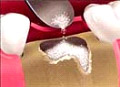
Detaching mucous-periosteum flasp creates an access to bony hole where tooth was removed.
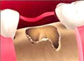
The preparation of the artificial bone is packed in the hole that was previously cleaned and freshened.
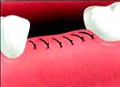
For a better wound healing and integration of bony tissue the frame is tightly sewn.
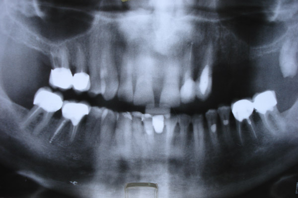
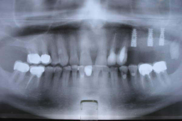
4-6 months later since complete bone integration, the doctor will make a panoramic x-ray picture of jawbones and will decide upon the subsequent implantation.
These and other technologies are used in the dental centre “Denta Vita” located in the centre of Chisinau, where our qualified dentists take advantage of all their experience with the help of the up-to-date equipment for our patients’ benefit.